Open this record in the complete research map
Cell Death in the Epithelia of the Intestine and Hepatopancreas in Neocaridina heteropoda (Crustacea, Malacostraca)
Sonakowska et al., 2016. PLOS ONE 11(2): e0147582.
This record is a claim boundary, not a quality badge. Evidence class and species relationship describe what was studied. They do not make the result universal, complete or automatically applicable to a home aquarium.
- Source taxon as published
- Neocaridina heteropoda obtained from local breeders; no host-identification method reported
- Environment
- Adults maintained in 30 L laboratory aquaria at 24 C, pH 7 and 15 degrees German total hardness and fed a commercial freshwater-shrimp food
- Life stages
- adult male, adult female
- Reviewed
- 2026-08-12
What was studied
Study shape: Light, transmission-electron and confocal microscopy, acid-phosphatase and LysoTracker staining, TUNEL labeling and JC-1 flow cytometry were used to describe autophagy, apoptosis, necrosis and mitochondrial potential in intestine and hepatopancreas tissue from method-specific adult groups. No external stressor or disease treatment was assigned.
Experimental unit: Prepared tissue or cell suspension from method-specific adult specimens: 15 females and 15 males for general microscopy, three adults for quantitative necrosis and autophagy cell counts, two for TEM acid-phosphatase staining, 10 females and 10 males for cryosections, five pooled within each organ suspension preparation and seven for JC-1 flow cytometry; the TUNEL animal denominator was not stated
Endpoints: cell-type and organ location, autophagic structures, TUNEL-positive apoptotic cells, necrotic-cell morphology, mitochondrial membrane potential, sex comparison.
Claim boundary
What it can support: Under the described laboratory history and without an assigned external stressor, the examined adult tissues contained autophagic, apoptotic and occasional necrotic cells. Autophagy was observed in mature intestine D cells and hepatopancreas B and F cells, apoptosis in anterior-intestine D cells and proximal hepatopancreas B and F cells, while regenerative cells showed none of the three reported processes. The study did not report a statistically significant intestine-versus-hepatopancreas difference in cells with depolarized mitochondria.
What it cannot support: This is destructive cellular and tissue evidence, not a visible home diagnosis or a disease experiment. Breeder source, sex and adult status were reported, but host identification, age, molt stage, reproductive state, tank count, animals per tank, acclimation duration, health screening, water-test methods and the TUNEL animal denominator were not. Method groups differed and their overlap was not reported. Cell counts were nested within only three adults for the necrosis and autophagy percentages, TUNEL used an arbitrary fluorescence threshold and sex-specific values or tests were not shown. Table 1 prints 938 autophagic cells from 231 total for intestine specimen 3 even though the displayed 40.26 percent implies 93. The results do not establish a gross sign, pathogen, cause of illness, welfare state, treatment target, aquarium optimum or safe exposure, and the maintained 24 C, pH 7 and 15 degree hardness history was not an assigned comparison.
Publication status audit
Matched in dated Crossref query
The canonical DOI appeared in the dated broad query and carried no registered update in that response. This is a metadata observation, not proof that the work has never changed.
Metadata snapshot reviewed 2026-08-12. Inspect the method, unmatched queue and limitations.
Correction impact
If this source boundary changes, these are the known downstream consumers. The list distinguishes scientific re-review from generated parity and historical follow-up.
Editorial review required (4)
This curated wording or synthesis must be reconsidered when the source boundary changes. A passing generator test cannot decide the new scientific meaning.
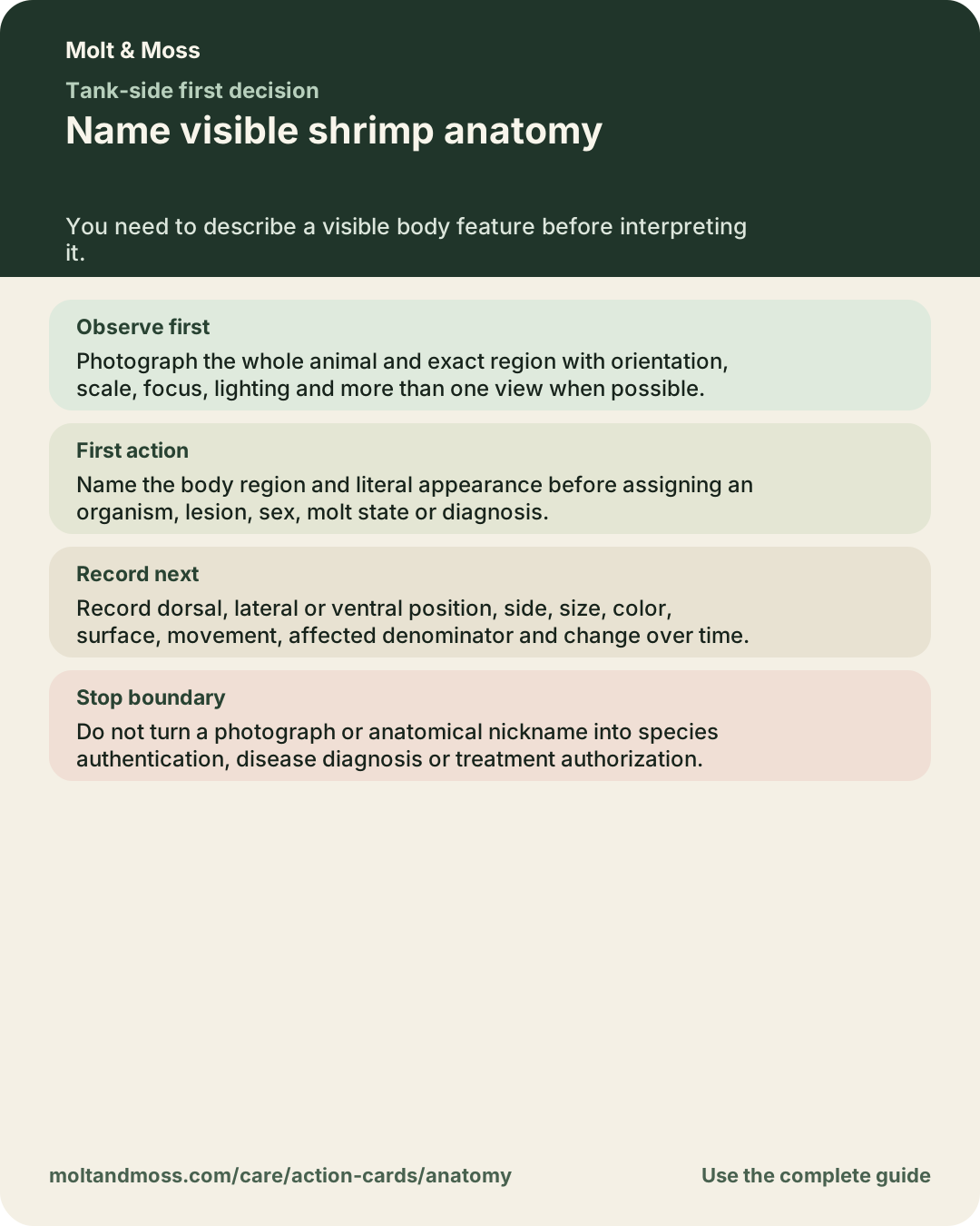
- anatomy decision card Tank-side decision card
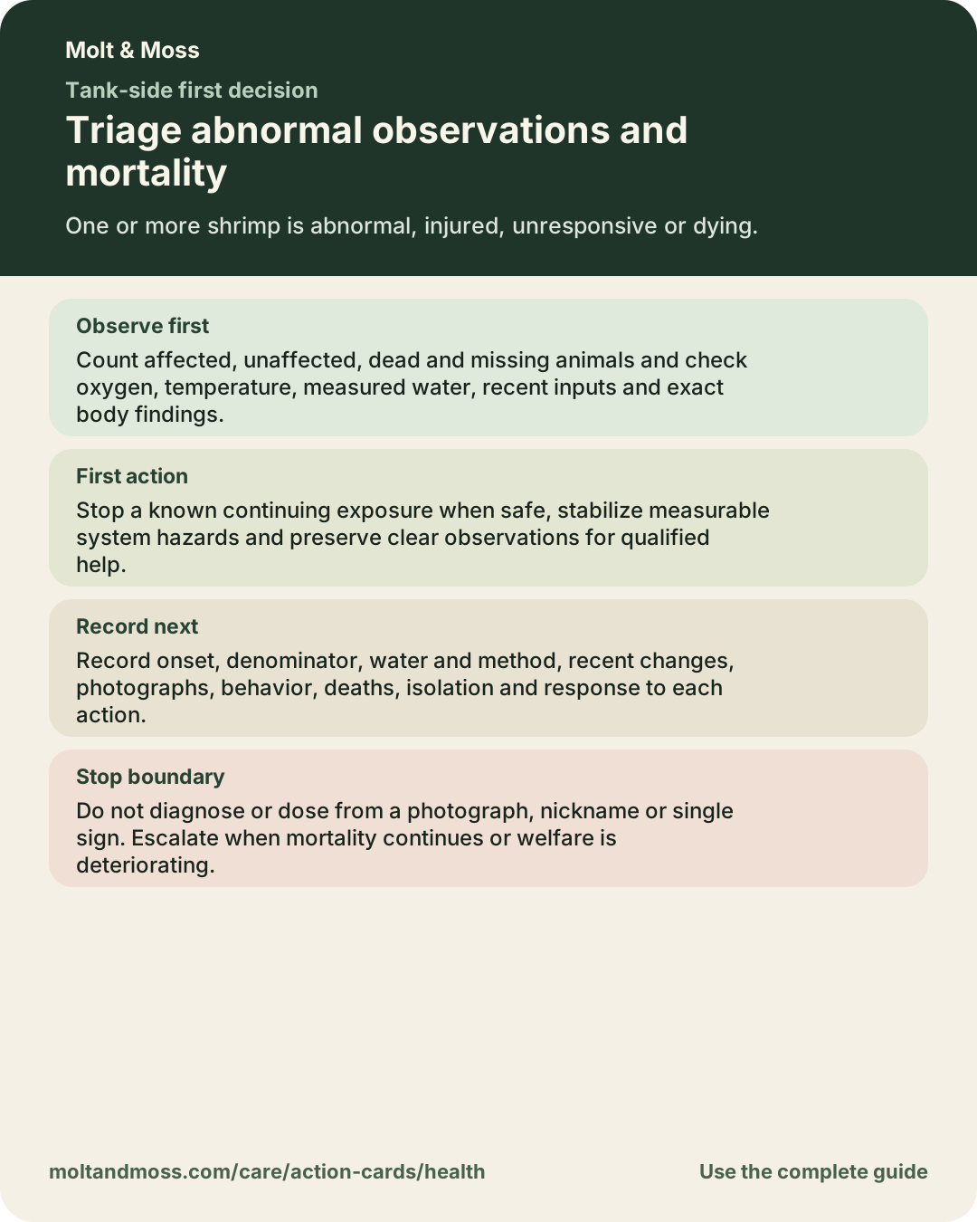
The card translates this guide into an observation, first action, next record and stopping boundary, so all four require editorial review when the source changes. - health decision card Tank-side decision card
The card translates this guide into an observation, first action, next record and stopping boundary, so all four require editorial review when the source changes. - anatomy practical guide Practical guide
The guide uses this evidence record and its practical wording must remain inside the revised source boundary. - health practical guide Practical guide
The guide uses this evidence record and its practical wording must remain inside the revised source boundary.
Generated from governing registry (13)
This surface derives from a governing registry and should update with it, but release tests must still prove parity and routing.
- Record CSL-JSON citation Citation export
The record citation export derives its note and source metadata from the evidence registry. - Record RIS citation Citation export
The record citation export derives its note and source metadata from the evidence registry. - Complete CSL-JSON bibliography Citation export
The complete citation export includes every evidence registry record. - Complete RIS bibliography Citation export
The complete citation export includes every evidence registry record. - anatomy portable decision card Generated decision-card text
The portable text derives from the curated decision-card registry and must preserve every card section and both permanent links. - health portable decision card Generated decision-card text
The portable text derives from the curated decision-card registry and must preserve every card section and both permanent links. - anatomy complete visual decision card Generated decision-card visual
The typography-only visual and exact accessibility description derive from the curated decision-card registry and must preserve every card section. - health complete visual decision card Generated decision-card visual
The typography-only visual and exact accessibility description derive from the curated decision-card registry and must preserve every card section. - Cell Death in the Epithelia of the Intestine and Hepatopancreas in Neocaridina heteropoda (Crustacea, Malacostraca) Canonical evidence page
The canonical evidence page derives from the current evidence registry record. - anatomy evidence dossier Generated guide dossier
The dossier derives its linked evidence set from the record care path. - health evidence dossier Generated guide dossier
The dossier derives its linked evidence set from the record care path. - Machine-readable knowledge export Machine-readable knowledge export
The versioned export includes the complete evidence registry and source-integrity audit. - Research map record Research map
The research map derives its source boundary from the evidence registry.
Historical record needs follow-up decision (1)
Do not silently rewrite history. Decide whether the prior change record remains accurate and publish a new correction or scope note when needed.
- A cell-death marker is not automatically a shrimp disease Historical research change
The historical change cites this source. Preserve the old decision and decide whether a new public correction is required.
How this record is classified
Morphology or taxonomy: Anatomy, specimen identification or taxonomic relationships were studied with stated methods. A trade name or photograph cannot replace those methods.
Direct Neocaridina evidence: The source measured animals named within Neocaridina. Direct still does not mean universal or sufficient.
Critically appraise this source without a score
These questions fit this record's morphology or taxonomy role. They are prompts, not automatic judgments. Open the original source to answer them.
- Identity and provenance: How were the organism, population, stock and life stage identified and sourced? A published name or seller label can hide a taxonomic, population or life-stage transfer.
- Independent unit: What unit was independently assigned or sampled, and how many units support each comparison? Animal counts do not create independent replication when animals share a vessel, site, family or treatment history.
- Comparison and context: What was the actual comparator, allocation process, environment, duration and material condition? A result has meaning only against the comparison and conditions that produced it.
- Endpoint and measurement: Which endpoint was measured, with what method, unit, timing, resolution and decision rule? One endpoint cannot silently become survival, welfare, diagnosis, reproduction or long-term population performance.
- Denominators and missingness: Are starting counts, exclusions, losses, missing observations and analysis denominators reconciled? Unreported or changing denominators can alter the apparent direction, precision and applicability of a result.
- Transfer boundary: What is the nearest tempting aquarium claim that this source design cannot establish? Direct evidence can still be narrow, and adjacent evidence can be useful only while the inference remains visible.
- Examined material: Which specimens, localities, sexes, stages, types or sequence accessions were actually examined? A diagnosis applies to examined material and methods, not automatically to a trade-line photograph.
- Diagnostic characters: Which characters or loci distinguish the taxon, and were alternatives explicitly compared? Color alone may be variable, convergent or unrelated to the diagnostic character.
- Name history: Were synonyms, historical combinations, type material and later revisions checked? An older title can describe a population that later work placed in another taxon.
- Consumer identification limit: Can the required character or method be observed reliably in an ordinary live-animal image? A valid taxonomic method may still be unavailable to a keeper examining one photograph.
Where it is used
Reuse the reviewed citation boundary
Download CSL-JSON or download RIS. Both retain this record's support, limitation and review note.
Challenge a transcription, interpretation or missing boundary. A challenge changes the record only after editorial review.